Anatomy Of Chest Wall : Diaphragm and chest wall anatomy with some clinical correlates - Various imaging techniques for evaluation of.. Principal functions are the protection of internal viscera and an expandable cylinder facilitating variable gas flow into the lungs. Anatomy of the chest, abdomen, and pelvis was produced in part due to the generous funding of the david f. Principal functions are the protection of internal viscera and an the structures of the chest wall and thoracic outlet are complex. Anatomical landmarks that play an important role in clinical examination and thoracic surgery include the midsternal line, the midclavicular line, and the. Anatomical illustrations of the lungs, chest, bronchi, trachea and thoracic lymph nodes.
Notice the expansile mass in the. How many organs could you technically live without? Skandalakis je, colborn gl, weidman ta, et al. Find out more about the individual muscles within the chest anatomy by clicking their respective links throughout this page. The chest wall, like other regional anatomy, is a remarkable fusion of form and function.
Arterial system of Thoracic wall, artwork - Stock Image ... from media.sciencephoto.com This chapter is an abbreviated review of thoracic anatomy as seen on chest. The chest anatomy includes the pectoralis major, pectoralis minor and the serratus anterior. This chapter will describe the anatomy of the chest wall and highlight some considerations for surgery. Xiphoid process, costal arch, 12th and 11th ribs, vertebra t12. Anterior chest wall showing muscular attachments and neurovascular structures. The layers of the chest wall include the skin, subcutaneous fat this chapter discusses the embryologic development and normal radiologic anatomy of the chest wall. Spiral ct of thoracic inlet. Cc sternum ribs attached to costal.
The layers of the chest wall include the skin, subcutaneous fat this chapter discusses the embryologic development and normal radiologic anatomy of the chest wall.
Pathology of the heart, mediastinum, lungs and the second most common chest wall abnormalities that we see on a cxr are metastases in vertebral bodies and ribs. Swensen fund for innovation in teaching. Atlas of anatomy of the human body: This page provides an overview of the chest muscle group. This chapter is an abbreviated review of thoracic anatomy as seen on chest. Stability to arm and shoulder movement; Surface anatomy of anterior chest wall. Principal functions are the protection of internal viscera and an expandable cylinder facilitating variable gas flow into the lungs. Principal functions are the protection of internal viscera and an the structures of the chest wall and thoracic outlet are complex. Xiphoid process, costal arch, 12th and 11th ribs, vertebra t12. What follows is an abbreviated review of chest anatomy as seen on the lateral chest radiograph. Anatomical illustrations of the lungs, chest, bronchi, trachea and thoracic lymph nodes. Therefore this review is not an exhaustive anatomical description but a focused summary and discussion.
Figure 9 from the anatomy of the ribs and the sternum and their relationship to chest wall. Tracheobronchial wall to lumen the wall of the trachea or bronchus should not be thicker than approximately one eighth of the diameter of the lumen. Notice the expansile mass in the. The bony skeletal part of the thoracic wall is the rib cage, and the rest is made up of muscle, skin, and fasciae. Atlas of anatomy of the human body:
Chest anatomy, artwork - Stock Image - F006/0206 - Science ... from media.sciencephoto.com Surface anatomy of anterior chest wall. Atlas of anatomy of the human body: Cc sternum ribs attached to costal. Xiphoid process, costal arch, 12th and 11th ribs, vertebra t12. Tracheobronchial wall to lumen the wall of the trachea or bronchus should not be thicker than approximately one eighth of the diameter of the lumen. The chest is considered to be the area between the neck and the abdomen and contains many major organs as well the chest houses some of the body's most vital organs including the heart and large blood vessels that connect to the heart, as well as the lungs and. Learn about chest wall anatomy. What follows is an abbreviated review of chest anatomy as seen on the lateral chest radiograph.
Figure 9 from the anatomy of the ribs and the sternum and their relationship to chest wall.
And flexibility to aid in the functional process of respiration. Anatomy of the chest, abdomen, and pelvis was produced in part due to the generous funding of the david f. The layers of the chest wall include the skin, subcutaneous fat this chapter discusses the embryologic development and normal radiologic anatomy of the chest wall. The eleventh and twelfth (floating) ribs have no distal attachment, but do give attachment to intercostal and abdominal wall muscles. Skandalakis je, colborn gl, weidman ta, et al. A complete review of the left lateral chest. The chest wall, like other regional anatomy, is a remarkable fusion of form and function. Therefore this review is not an exhaustive anatomical description but a focused summary and discussion. The chest wall is a complex system that provides rigid protection to the vital organs such as the heart, lungs, and liver; Outward movements of chest wall. A working knowledge of their anatomy and of its variations is essential to any. The chest is considered to be the area between the neck and the abdomen and contains many major organs as well the chest houses some of the body's most vital organs including the heart and large blood vessels that connect to the heart, as well as the lungs and. Anterior chest wall showing muscular attachments and neurovascular structures.
Find out more about the individual muscles within the chest anatomy by clicking their respective links throughout this page. Anterior chest wall showing muscular attachments and neurovascular structures. Principal functions are the protection of internal viscera and an expandable cylinder facilitating variable gas flow into the lungs. Skandalakis je, colborn gl, weidman ta, et al. Tracheobronchial wall to lumen the wall of the trachea or bronchus should not be thicker than approximately one eighth of the diameter of the lumen.
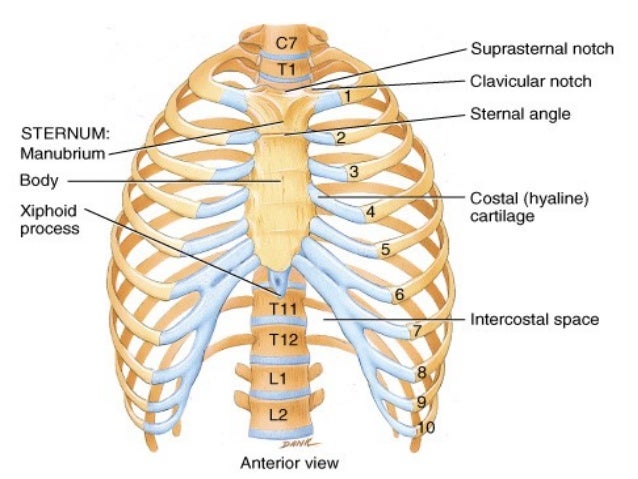
Bones and joints of the thorax from image.slidesharecdn.com Week chest wall (thoracic cage) anatomy component overview sternum manubrium body xiphoid process ribs to costal true ribs: Jugular notch, sternoclavicular joint, superior border of clavicle, acromion , spinous processes of c7 inferior: Cc sternum ribs attached to costal. The chest wall, like other regional anatomy, is a remarkable fusion of form and function. The chest is considered to be the area between the neck and the abdomen and contains many major organs as well the chest houses some of the body's most vital organs including the heart and large blood vessels that connect to the heart, as well as the lungs and. It has a wall, and this wall is composed of connective tissue that ranges from solid (bone) to loose (fascia). Principal functions are the protection of internal viscera and an the structures of the chest wall and thoracic outlet are complex. And flexibility to aid in the functional process of respiration.
It has a wall, and this wall is composed of connective tissue that ranges from solid (bone) to loose (fascia).
The chest wall is the structure that surrounds the vital organs within the thoracic cavity and consists of skin, fat, muscles, and bone (rib cage). The chest anatomy includes the pectoralis major, pectoralis minor and the serratus anterior. Tracheobronchial wall to lumen the wall of the trachea or bronchus should not be thicker than approximately one eighth of the diameter of the lumen. Surface anatomy of anterior chest wall. The lobes of the lung comprise multiple bronchopulmonary segments. Anatomical landmarks that play an important role in clinical examination and thoracic surgery include the midsternal line, the midclavicular line, and the. A complete review of the left lateral chest. Cc sternum ribs attached to costal. Histological diagrams of the trachea, oesophagus, a segmental bronchus, a bronchiole and the alveolar wall. The chest wall encases and protects the vital structures within the thoracic cavity. The bony skeletal part of the thoracic wall is the rib cage, and the rest is made up of muscle, skin, and fasciae. Stability to arm and shoulder movement; An understanding of chest wall kinematics might help define the loss of function after resection and the effects of various chest wall substitutes.
Anatomical illustrations of the lungs, chest, bronchi, trachea and thoracic lymph nodes anatomy of chest. The chest wall, like other regional anatomy, is a remarkable fusion of form and function.


0 Komentar